
How Brain Injuries Are Diagnosed
According to the CDC, it is estimated that 1.5 million Americans sustain a traumatic brain injury (TBI) each year. Motor vehicle crashes are the leading cause of hospitalization for TBIs. When someone has been in a motor vehicle collision, or in an incident where there has been contact to the head or a whiplash-type event, it is important for that person to be evaluated for a TBI.
When evaluating someone for a TBI, first responders and doctors typically start with the Glasgow Coma Scale. This is a 15-point test that assesses “impairment of consciousness” that looks at eye opening response, verbal response, and motor response. Points are given for each part of the test and added up. Injuries are then classified based as follows:
Severe – score of 8 or less
Moderate – score of 9 to 12
Mild – score of 13 to 15
But just because someone has a score between 13-15 does not mean they did not suffer a serious brain injury. The Glasgow Coma Score really just measures how conscious a person is following a suspected TBI. We have had many clients with brain injuries that caused lifelong complications who also had a 15 Glasgow Coma Score in the emergency room.
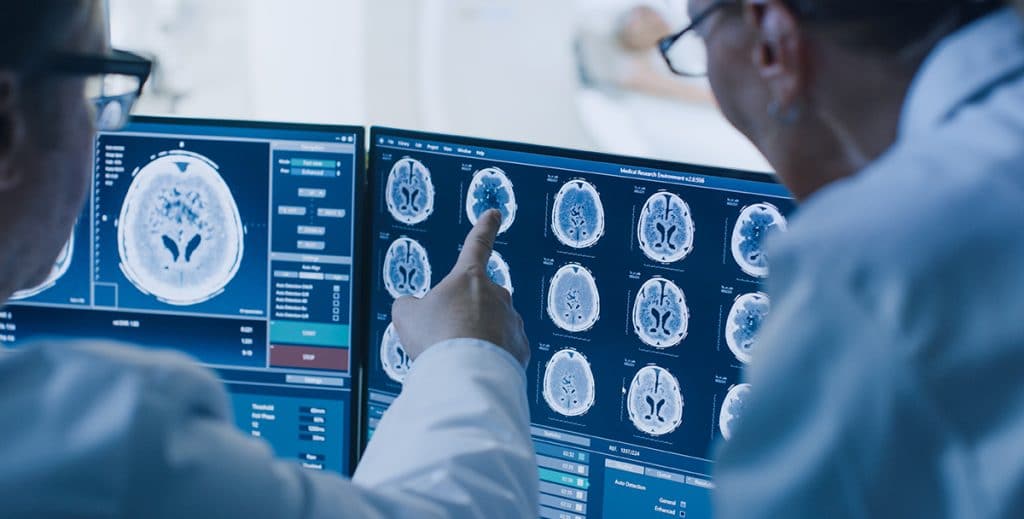
If further evaluation is needed, doctors turn to different types of medical imaging. In emergency rooms, doctors are most concerned with looking for serious injuries that need to be treated immediately. Doctors will usually start with CT scans to look for things like skull fractures, swelling of the brain, bleeding in the brain, and blood clots.
CT scans are made up a series of x-rays and will not show a detailed view of the brain. Because of this, a normal CT scan does not necessarily mean that the person does not have a brain injury. Again, the primary concern is looking for injuries that need to be treated immediately in the emergency room, not at detailed images of the brain.
If doctors suspect a brain injury that cannot be seen on a CT scan they will then turn to MRIs. Instead of using x-rays, MRIs use a very strong magnet and radio waves to look at protons in water in the brain. Since our brains have a lot of water in the soft tissue, MRIs are useful in showing the structure of the brain.
MRI images are divided into slices about a quarter inch thick, which allows doctors to view different layers of the brain. Think of it like slicing a loaf of bread then being able to look at each slice of bread from top, bottom, back, front, or sides.
On the MRI image, different parts of the brain will appear white, black, or gray. This depends on things like amounts of fat and water in the brain tissue. Things like air and bone will appear black, while soft tissue, blood, and other fluids will vary from white to black. The doctor reading the image looks at the differences between white and black areas to figure out if tissue in the brain is healthy or not. MRIs are useful for seeing things like strokes, aneurysms, bleeding, and tumors.
MRIs can also be used to show a type of brain injury called diffuse axonal injuries. Quick detour from imaging while we explain diffuse axonal injuries. Diffuse axonal injuries are injuries that happen when the brain shifts rapidly in the skull causing tearing of nerve fibers (axons) that connect different parts of the brain and send messages to the different parts of the brain. This can happen from a blow to the head or rapid movement of the head that causes the brain to move violently around and hit the skull.
These injuries can show up as lesions in MRI images and are many times located in the gray-white matter junction of the brain. Gray matter is generally made up of cell bodies while white matter is made up of axons. Axons are what connect brain cells and allow them to communicate with each other. When the axons tear, it shows up on the MRI.
While diffuse axonal injuries can show up on MRIs, technology now allows doctors to see how those injuries have impacted the brain. Functional MRI (fMRI) and diffusion tensor imaging (DTI) allow doctors to measure how blood and water move in the brain to evaluate brain activity.
fMRI shows which parts of the brain are activated when certain functions are performed. It does this by measuring changes in blood flow while the person alternates between a resting state and performing certain tasks. The results show which parts of the brain handle different functions and which parts are no longer functioning as they should.
DTI also uses MRI technology and looks at how and in what direction water molecules move along axonal tracts. When the brain suffers a diffuse axonal injury the axonal tracts become damaged and the signals that normally move along these paths become interrupted. DTI is able to show this interruption by showing the interruption in the travel of water molecules.
Finally, electroencephalograms (EEG) are sometimes used to diagnose brain injuries, though used less frequently than other imaging techniques. EEGs are used to detect electrical activity in the brain and are commonly used to detect epilepsy and seizures, but can also be used to detect stroke or lack of brain activity in someone who is in a coma.
Brain injuries are serious injuries and can have lifelong effects on the injured person and their family. Not all lawyers understand brain injuries and even fewer have taken brain injury cases to trial. If you, or someone in your family, has suffered a traumatic brain injury it’s important to research lawyers and contact a lawyer with experience in handling brain injuries. Contact the Las Vegas brain injury attorneys at Claggett & Sykes at 702-333-7777, or fill out our contact form to schedule a free consultation. You can also schedule an appointment in our Reno offices.

We are not simply a personal injury firm. We are trial lawyers who take on catastrophic injury, brain injury, and wrongful death cases. These cases are different than most personal injury cases and the needs of these cases cannot be met by law firms that take on just any case.
Read more about Claggett & Sykes Trial Lawyers